Understand the structure of the heart Help prevent cardiovascular disease


Although the heart is only the size of a fist, it is the center of the lifeblood and the core of the human blood circulation system. It is responsible for transporting blood to various organs throughout the body. You can imagine how complex the structure is. Let us understand the structure of the heart in the following article!
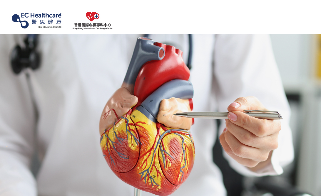
The heart is a hollow organ made of muscle tissue located between the lungs on the left side of the chest. It is basically composed of 4 parts: atrium, ventricle, heart valves and heart blood vessels:
1. Atria: located in the upper part of the heart.
.Left atrium: receives oxygenated blood from the lungs.
.Right atrium: receives deoxygenated blood from veins throughout the body.
2. Ventricles: Located in the lower part of the heart.
.Left ventricle: Thick muscle that pumps oxygenated blood throughout the body.
.Right ventricle: Thin muscle that pumps only deoxygenated blood to the lungs.
3. Heart valve: It is the valve structure between the atrium and ventricle as well as the ventricle and aorta. It is responsible for controlling the single flow direction of blood during cardiac contraction and relaxation to prevent reverse flow.
.Mitral valve: Located between the left atrium and left ventricle, it prevents blood from flowing back into the left atrium.
.Tricuspid valve: Located between the right atrium and right ventricle, it prevents blood from flowing back into the right atrium.
.Aortic valve: Located between the left ventricle and the aorta, it prevents blood from flowing back into the left ventricle.
.Pulmonary valve: Located between the right ventricle and the pulmonary artery, it prevents blood from flowing back into the right ventricle.
4. Heart blood vessels: Arteries pump oxygenated blood from the heart to organs and tissues, while veins carry deoxygenated blood containing metabolic waste products from organs and tissues back to the heart. The aorta, superior and inferior vena cava, pulmonary artery and pulmonary veins form the systemic circulation and pulmonary circulation, forming a complete circuit of blood circulation.
.Coronary Arteries: The left and right coronary arteries surround the heart, supplying oxygen and nutrients to the heart and maintaining normal heart function.
.Aorta: Also known as systemic circulation artery, starting from the left ventricle, it transports oxygenated blood to the whole body.
.Superior and inferior vena cava: Deoxygenated blood from the whole body is transported to the right atrium. The superior vena cava collects blood from the upper body, and the inferior vena cava collects blood from the lower body.
.Pulmonary artery: starts from the right ventricle, it carries deoxygenated blood to the lungs.
. Pulmonary veins: start in the lungs, it carries oxygenated blood to the left atrium.
The heart muscle is composed of three layers: pericardium, myocardium and endocardium. The pericardium is the outermost membrane surrounding the heart, which protects and stabilizes the heart and reduces friction. Myocardium is the muscle layer of the heart, responsible for cardiac contraction and relaxation functions, and can also secrete a variety of other biologically active substances, such as brain natriuretic peptide, renin, etc. The endocardium is the innermost layer of the heart, covering the heart valves and the smooth surface of the four heart chambers. When blood flows in the heart chambers, it can reduce frictional resistance and prevent blood coagulation.
Although the structure of the heart is quite complex, having a basic understanding of it will help us better understand the induction mechanism of cardiovascular diseases and further prevent or treat related diseases.
Related Brands


